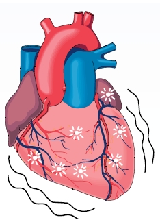
Tuesday, June 23, 2009

Bronchopulmonary Dysplasia


What Is Bronchopulmonary Dysplasia?


Bronchopulmonary dysplasia, or BPD, is a serious lung condition that affects mostly babies who:


  • Are born more than 10 weeks before their due dates


  • Weigh less than 2½ pounds, or 1,000 grams, at birth


  • Have breathing problems at birth


  • Need long-term breathing support and oxygen


Many of these babies are born with serious respiratory distress syndrome (RDS). Their lungs haven't yet developed enough to make surfactant (sur-FAK-tant). Surfactant is a liquid that coats the inside of the lungs and keeps them open so that the baby can breathe in air once he or she is born.


As a result, these babies are usually put on oxygen and a breathing machine at birth—either a ventilator (also known as a respirator) or a nasal continuous positive airway pressure (NCPAP) machine. This can prevent damage to their brains and other body organs from lack of oxygen. They also are given surfactant.


Most babies with RDS begin to get better within the next 2 to 4 weeks. But some get worse and need more oxygen and/or breathing assistance from a machine. These babies have developed BPD.


The lungs of the babies who are born with RDS and go on to develop BPD are less developed than those of babies with RDS who recover. They usually have fewer and larger alveoli, or air sacs, than other newborns. They also may have fewer tiny blood vessels in the alveoli. The blood vessels are needed to move oxygen from the alveoli into the bloodstream.


These babies also are more likely than other infants to have problems in other parts of their bodies that aren't yet fully developed. These include the heart, kidneys, brain, stomach, intestines, and eyes.


With new and better treatments now available, most babies with BPD get better over time, and many go on to live normal, active lives.


What Causes Bronchopulmonary Dysplasia?


The lungs of babies born more than 10 weeks before they are due are fragile and easily irritated or injured by things in the outside environment during the first hours or days after birth.


Doctors now believe that a baby gets bronchopulmonary dysplasia (BPD) as a result of the way his or her lungs respond to some of these things, including:


  • High levels of oxygen. Doctors usually give oxygen to newborns with breathing problems. This is to make sure that their brains, hearts, livers, and kidneys receive enough oxygen to do their jobs. But high levels of oxygen can cause inflammation in the lungs. This can result in injury to the breathing passages. High levels of oxygen also can slow the normal development of the lungs in babies born very early.


  • Pressure caused by mechanical ventilation. In the past, doctors usually put newborns who couldn't breathe on their own on mechanical ventilators. These machines apply pressure to push air into the babies' lungs. This pressure can irritate the lungs and cause them to become more inflamed. Mechanical ventilation is a factor in most cases of BPD.


  • Doctors try to minimize the injury by using ventilation only when absolutely needed. Today, more and more doctors are putting these babies on nasal continuous positive airway pressure (NCPAP) machines, which don't put the same kind of pressure on the babies' lungs.


  • Infections. Infections in babies born early can cause inflammation in their underdeveloped lungs. This narrows the breathing passages and makes it harder for the baby to breathe. Lung infections also increase the baby's need for extra oxygen and help with breathing.


Some doctors think that heredity may be a factor in the development of BPD.


What Are the Signs and Symptoms of Bronchopulmonary Dysplasia?


Most babies who get bronchopulmonary dysplasia (BPD) are born with respiratory distress syndrome (RDS). The signs and symptoms of RDS at birth are:


  • Rapid, shallow breathing


  • Sharp pulling in of the chest below the ribs with each breath taken in


  • Grunting sounds during exhalation


  • Flaring of the nostrils during breathing


As a result, these babies usually are put on a breathing machine right away. This is to prevent damage to their brains, hearts, and other body organs from lack of oxygen. These babies also are given surfactant to coat the tiny air sacs and to help prevent their lungs from collapsing.


Doctors can usually diagnose BPD after about 2 weeks. At this point, the baby hasn't started getting better, and he or she needs more oxygen.


Babies with severe BPD may also develop:


  • Pulmonary arterial hypertension, continuous high blood pressure in the blood vessels that carry oxygen-poor blood from the right ventricle in the heart to the small arteries in the lungs


  • Cor pulmonale, failure of the right side of the heart caused by ongoing high blood pressure in the pulmonary artery and right ventricle


How Is Bronchopulmonary Dysplasia Diagnosed?


It's hard to tell whether a baby with breathing problems has bronchopulmonary dysplasia (BPD) before he or she is about 14 to 30 days old. At this point, the baby should be showing improvement in the breathing problems. Instead, the baby's condition seems to be getting worse and he or she needs more oxygen or help from a breathing machine.


Doctors usually conduct a number of tests on newborns with breathing problems to make sure they diagnose their condition correctly. These tests include:


  • Blood tests. Blood samples are checked to see whether the baby has enough oxygen in his or her blood.


  • Chest x ray. A chest x ray takes a picture of the heart and lungs. It shows larger areas of air and changes from inflammation or infection. It also shows areas of the lung that have collapsed and may help confirm that the lungs aren't developing normally.


  • Echocardiogram. This test uses sound waves to create a moving picture of the heart. Echocardiogram is used to rule out congenital heart defects orpulmonary arterial hypertension as the cause of the breathing problems.


Doctors grade BPD as mild, moderate, or severe, depending on how much extra oxygen the baby needs and how long he or she needs it.


How Can Bronchopulmonary Dysplasia Be Prevented?


You can do certain things to help ensure that your baby isn't born before his or her lungs have developed completely.


They include:


  • Seeing your doctor regularly during your pregnancy


  • Eating right


  • Avoiding tobacco smoke, alcohol, and illegal drugs


  • Controlling any ongoing medical conditions you have


  • Preventing infection


Your doctor may also recommend that you take progesterone if you had a prior preterm birth. This is a hormone that may help delay delivery.


Your doctor may give you injections of a corticosteroid medicine if it looks as though you may give birth too early. This medicine can speed up surfactant production and development of the lungs, brain, and kidneys in the fetus. Usually, within about 24 hours after you start taking the medicine, the fetus' lungs will work better. They also will respond better to surfactant treatment so that respiratory distress syndrome will not develop after delivery, or it will be relatively mild.


How Is Bronchopulmonary Dysplasia Treated?


The goals of treatment for babies with bronchopulmonary dysplasia (BPD) are to:


  • Reduce further injury to the lungs


  • Provide nutrition and other support to help the lungs grow and recover


Treatment is done in three stages. They are:


  • Treatment for respiratory distress at birth and before doctors know whether the baby has BPD


  • Treatment after doctors know the baby has BPD


  • Home care after the baby leaves the hospital


Treatment of respiratory distress usually begins as soon as the baby is born, sometimes in the delivery room. Most infants who show signs and symptoms ofrespiratory distress syndrome (RDS) are quickly moved to a special intensive care unit called a neonatal intensive care unit (NICU). There they receive around-the-clock treatment from a group of health care professionals who specialize in treating premature infants.


The most important treatments for RDS are:


  • Breathing support


  • Surfactant replacement therapy


Breathing Support


These babies usually are put on a breathing machine to help them breathe—either a mechanical ventilator or a nasal continuous positive airway pressure (NCPAP) machine. The ventilator is connected to a breathing tube that runs through the baby's mouth or nose into the windpipe. The ventilator can be set to help a baby breathe or to completely control a baby's breathing. It also is set to give the amount of oxygen the baby needs.


Today, more and more babies are receiving breathing support from an NCPAP machine, which pushes air into the baby's lungs through prongs in the nostrils.


With breathing help, the baby's lungs have a chance to develop. Breathing machines today don't cause as much injury to the airways and lungs as those used in the past.


Surfactant Replacement Therapy


The baby is given surfactant to open his or her lungs until the lungs have developed enough to start making their own surfactant. Surfactant is given through a tube that is attached to the breathing machine, which pushes the surfactant directly into the baby's lungs.


Other Types of Treatment


Other treatments for babies who show signs and symptoms of RDS and haven't yet been diagnosed with BPD include:


Medicines


Doctors usually give the baby medicines to reduce swelling in the airways and improve the flow of air in and out of the lungs. These medicines include:


  • Bronchodilators to improve the flow of air in and out of the lungs


  • Diuretics to help remove extra fluid from the lungs


  • Antibiotics to control infections


Supportive Therapy


Treatment in the NICU is designed to limit stress on the baby and meet his or her basic needs of warmth, nutrition, and protection. Such treatment usually includes:


  • Using a radiant warmer or incubator to keep your baby warm and reduce the chances of infection.


  • Ongoing monitoring of blood pressure, heart rate, breathing, and temperature through sensors taped to the baby's body.


  • Using a sensor on a finger or toe to monitor the amount of oxygen in the baby's blood.


  • Giving fluids and nutrients through a needle or tube inserted into a vein to prevent malnutrition and promote growth. Nutrition is critical to the growth and development of the lungs. Later, your baby may be given milk through a tube that is passed through his or her nose into the mouth.


  • Monitoring fluid intake to make sure that fluid doesn't build up in the baby's lungs.


Once doctors know that the baby has BPD, some or all of these treatments are continued in the NICU:


  • Babies with BPD are usually taken off the breathing machine slowly over time. They are often moved from the ventilator to an NCPAP machine until they can breathe on their own. This machine pushes air into the baby's lungs through prongs in the nostrils.


  • The baby is likely to continue to need extra oxygen for some time. Once the baby no longer needs help breathing, he or she may get additional oxygen through prongs in the nostrils.


  • Babies with moderate to severe BPD have an echocardiogram every 2 to 3 months to check their pulmonary artery pressure.


These babies also may need physical therapy to strengthen their muscles and help their lungs clear out mucus.


Today, most babies with BPD recover. They may spend several weeks or months in the hospital. But the best place for the baby's growth and development is at home with the family where he or she can be in a loving and familiar environment.


After the baby goes home:


  • It's important for the parents to know about the symptoms and treatments for BPD. Parents and family members play an important role by being loving and involved with their babies and giving care.


  • The baby may continue to have some breathing symptoms and may remain in poor health.


  • He or she may still need extra oxygen and a breathing machine.


  • He or she needs good nutrition and extra calories because of the extra work involved with breathing.


  • Regular checkups and timely vaccinations from a pediatrician (a doctor who specializes in treating children) are important. A pediatrician also can treat any other illnesses that the baby may develop.


  • The family also may need social services to help them take care of the baby's medical and nonmedical needs.


What Is Sudden Cardiac Arrest (SCA)?


Sudden cardiac arrest (SCA) is acondition in which the heart suddenly and unexpectedly stops beating. When this happens, blood stops flowing to the brain and other vital organs.

SCA usually causes death if it's not treated within minutes.

Outlook

Ninety-five percent of people who have SCA die from it—most within minutes. Rapid treatment of SCA with a defibrillator can be lifesaving. A defibrillator is a device that sends an electric shock to the heart to try to restore its normal rhythm.

Automated external defibrillators (AEDs), which often are found in public places like airports and office buildings, can be used by bystanders to save the lives of people who are having SCA.

Causes

Most cases of sudden cardiac arrest (SCA) are due to ventricular fibrillation (v-fib). V-fib is a type of arrhythmia (for this please see my article on Wolff-Parkinson-White Syndrome (Arrhythmia). In v-fib, the ventricles (the heart's lower chambers) don't beat normally. Instead, they quiver very rapidly and irregularly.

When this happens, the heart pumps little or no blood to the body. V-fib is fatal if not treated within a few minutes.

Other electrical problems in the heart also can cause SCA. For example, SCA can occur if the rate of the heart's electrical signals becomes very slow and stops. SCA also can occur if the heart muscle doesn't respond to the heart's electrical signals.

Several factors can cause the electrical problems that lead to SCA. These factors include:

  • Coronary artery disease (CAD), which reduces blood flow to the heart muscle
  • Severe physical stress, which raises the risk for abnormal electrical activity in the heart
  • Inherited disorders that disrupt the heart's electrical activity
  • Structural changes in the heart that cause electrical signals to spread abnormally

Several research studies are under way to try to find the exact causes of SCA and how to prevent them.

Who Is At Risk?

Each year, between 250,000 and 450,000 Americans have sudden cardiac arrest (SCA). SCA occurs most often in people in their mid-thirties to mid-forties. It appears to affect men twice as often as women.

SCA rarely occurs in children unless they have inherited problems that make them likely to have SCA. Only a very small number of children have SCA each year.

Other Risk Factors

Other risk factors for SCA include:

  • A personal or family history of SCA or of inherited disorders that make you prone to arrhythmias
  • A history of having arrhythmias
  • Heart attack
  • Heart failure
  • Drug abuse or excessive alcohol intake

How Is Sudden Cardiac Arrest Diagnosed?

Sudden cardiac arrest (SCA) happens without warning. It requires immediate emergency treatment. Doctors rarely can diagnose SCA with medical tests as it's happening.

Instead, SCA often is diagnosed after it happens. Doctors do this by ruling out other causes of a person's sudden collapse.

How Is Sudden Cardiac Arrest Treated?

Emergency Treatment

Sudden cardiac arrest (SCA) requires immediate treatment with a defibrillator. This device sends an electric shock to the heart. The electric shock may restore a normal rhythm to a heart that's stopped beating.

To work well, defibrillation must be done within minutes of SCA. With every minute that passes, the chances of surviving SCA drop rapidly.

Police, emergency medical technicians, and other first responders usually are trained and equipped to use a defibrillator. Call 9–1–1 right away if someone has signs or symptoms of SCA. The sooner help is called, the sooner potentially lifesaving treatment can be done.

How Can Death Due to Sudden Cardiac Arrest Be Prevented?

Ways to prevent death due to sudden cardiac arrest (SCA) differ depending on whether:

  • You've already had SCA
  • You've never had SCA but are at high risk for the condition
  • You've never had SCA and have no known risk factors for the condition

For People Who Have Survived Sudden Cardiac Arrest

If you've already had SCA, you're at high risk of having it again. Research shows that an implantable cardioverter defibrillator (ICD) reduces the chances of dying from a second SCA.

An ICD is surgically placed under the skin in your chest or abdomen. The device has wires with electrodes on the ends that connect to your heart's chambers. The ICD monitors your heartbeat.

If the ICD detects a dangerous heart rhythm, it gives an electric shock to restore the heart's normal rhythm. Your doctor may give you medicine to limit irregular heartbeats that can trigger the ICD.

Implantable Cardioverter Defibrillator

The illustration shows the location of an implantable cardioverter defibrillator in the upper chest. The electrodes are inserted into the heart through a vein.

The illustration shows the location of an implantable cardioverter defibrillator in the upper chest. The electrodes are inserted into the heart through a vein.

An ICD isn't the same as a pacemaker. The devices are similar, but have some differences. Pacemakers only give off low-energy electrical pulses. They're often used to treat less dangerous heart rhythms, such as those that occur in the upper chambers of the heart. Most new ICDs work as both pacemakers and ICDs.

For People at High Risk for a First Sudden Cardiac Arrest

If you have severe coronary artery disease (CAD), you're at increased risk for SCA. This is especially true if you've recently had a heart attack.

Your doctor may prescribe a type of medicine called a beta blocker to help lower your risk for SCA. Other treatments for CAD, such as angioplasty or coronary artery bypass grafting, also may lower your risk for SCA.

Your doctor also may recommend an ICD if your risk for SCA is very high.

For People Who Have No Known Risk Factors for Sudden Cardiac Arrest

CAD seems to be the cause of most cases of SCA in adults. CAD also is a major risk factor for angina (chest pain or discomfort) and heart attack, and it contributes to other heart problems.

Following a healthy lifestyle can help you lower your risk for CAD, SCA, and other heart problems.

Healthy Diet and Physical Activity

A healthy diet is an important part of a heart healthy lifestyle. Choose a variety of fruits, vegetables, and grains; half of your grains should come from whole-grain products.

Choose foods that are low in saturated fat, trans fat, and cholesterol. Healthy choices include lean meats, poultry without skin, fish, beans, and fat-free or low-fat milk and milk products.

Choose and prepare foods with little sodium (salt). Too much salt can raise your risk for high blood pressure. Recent studies show that following the Dietary Approaches to Stop Hypertension (DASH) eating plan can lower blood pressure.

Choose foods and beverages that are low in added sugar. If you drink alcoholic beverages, do so in moderation.

Aim for a healthy weight by staying within your daily calorie needs. Balance the calories you take in with the calories you use while doing physical activity. Be as physically active as you can.

Some people should get medical advice before starting or increasing physical activity. For example, talk to your doctor if you have a chronic (ongoing) health problem, are on medicine, or have symptoms such as chest pain, shortness of breath, or dizziness. Your doctor can suggest types and amounts of physical activity that are safe for you.

For more information on following a healthy diet, see the National Heart, Lung, and Blood Institute's (NHLBI's) Aim for a Healthy Weight Web site, "Your Guide to a Healthy Heart," and "Your Guide to Lowering Your Blood Pressure With DASH." All of these resources provide general information about healthy eating.

For more information about physical activity, see NHLBI's "Your Guide to Physical Activity and Your Heart."

Other Lifestyle Changes

Other lifestyle changes also can help lower your risk for SCA. Examples include:

  • Quitting smoking. Talk to your doctor about programs and products that can help you quit. Also, try to avoid secondhand smoke.
  • Losing weight if you're overweight or obese.
  • Treating other health problems, such as high blood pressure, high blood cholesterol, and diabetes.

Wednesday, June 10, 2009

Bronchitis

What Is Bronchitis?

Bronchitis (bron-KI-tis) is a condition in which the bronchial tubes, the tubes that carry air to your lungs, become inflamed. (For more information on the bronchial tubes and the airways, see the Diseases and Conditions Index How the Lungs Work article.)

People who have bronchitis often have a cough that brings up mucus. Mucus is a slimy substance made by the lining of the bronchial tubes. Bronchitis also may cause wheezing (a whistling or squeaky sound when you breathe), chest pain or discomfort, a low fever, and shortness of breath.

Acute Bronchitis

Infections or other factors that irritate the lungs cause acute bronchitis. The same viruses that cause colds and the flu often cause acute bronchitis. These viruses are spread through the air when people cough. They also are spread through physical contact (for example, on hands that have not been washed). Sometimes bacteria cause acute bronchitis.

Acute bronchitis lasts from a few days to 10 days. However, the cough that occurs may last for several weeks after the infection is gone.

Several factors increase the risk for acute bronchitis. Examples include tobacco smoke (including secondhand smoke), air pollution, dust, and fumes. Avoiding these lung irritants as much as possible can help lower your risk for acute bronchitis.

Most cases of acute bronchitis go away within a few days. If you think you have acute bronchitis, see your doctor. He or she will want to rule out other, more serious health conditions that need medical care.

Chronic Bronchitis

Chronic bronchitis is an ongoing, serious condition. It occurs when the lining of the bronchial tubes is constantly irritated and inflamed.

Bronchitis is "chronic" if you have a cough with mucus on most days for at least 3 months a year and 2 years in a row (without another apparent cause). Smoking is the main cause of chronic bronchitis.

Viruses or bacteria can easily infect the irritated bronchial tubes. When this happens, the condition worsens and lasts longer. As a result, people who have chronic bronchitis also have periods when symptoms get much worse than usual.

Chronic bronchitis is a serious, long-term medical condition. Early diagnosis and treatment, combined with quitting cigarette smoking and avoiding secondhand cigarette smoke, can help people live better with this condition. The chance of complete recovery is low for people who have severe chronic bronchitis.

Symptoms of Bronchitis?

Acute Bronchitis

Acute bronchitis caused by an infection usually develops after you already have a cold or the flu. Symptoms of a cold or the flu include sore throat, fatigue (tiredness), fever, body aches, stuffy or runny nose, vomiting, and diarrhea.

The main symptom of acute bronchitis is a cough. The cough may produce clear mucus (a slimy substance). If the mucus is yellow or green, you may have a bacterial infection as well. Even after the infection clears up, you may still have a dry cough for days or weeks.

Other symptoms of acute bronchitis include wheezing (a whistling or squeaky sound when you breathe), low fever, and chest tightness or pain.

If your acute bronchitis is severe, you also may have shortness of breath, especially with physical activity.

Chronic Bronchitis

The signs and symptoms of chronic bronchitis include coughing, wheezing, and chest discomfort. The coughing may produce large amounts of mucus. This type of cough is often called "smoker's cough."

Diagnosis

Your doctor usually will diagnose bronchitis based on your signs and symptoms. He or she may ask questions about your cough, such as how long you’ve had it, what you’re coughing up, and how much you cough.

Your doctor also will likely ask:

  • About your medical history
  • Whether you’ve recently had a cold or the flu
  • Whether you smoke or spend time around others who smoke
  • Whether you’ve been exposed to air pollution, dust, or fumes

Your doctor will use a stethoscope to listen for wheezing (a whistling or squeaky sound when you breathe) or other abnormal sounds in your lungs. He or she also may:

  • Look at your mucus to see whether you have a bacterial infection
  • Test the oxygen levels in your blood using a sensor attached to your fingertip or toe
  • Order a chest x ray, lung function tests, or blood tests

What Causes Bronchitis?

Acute Bronchitis

Infections or other factors that irritate your lungs cause acute bronchitis. The same viruses that cause colds and the flu are the most common cause of acute bronchitis. Sometimes bacteria can cause the condition.

Certain substances can irritate your lungs and airways and raise your risk for acute bronchitis. For example, inhaling or being exposed to cigarette or cigar smoke, air pollution, dusts, vapors, or fumes raises your risk. These lung irritants also can make symptoms worse.

Being exposed to a high level of dust or fumes, such as from an explosion or a big fire, also may lead to acute bronchitis.

Chronic Bronchitis

Repeatedly breathing in fumes that irritate and damage lung and airway tissues causes chronic bronchitis. Smoking is the major cause of this condition.

Breathing in air pollution and dust or fumes from the environment or the workplace also can lead to chronic bronchitis.

People who have chronic bronchitis go through periods when symptoms become much worse than usual. During these times, they also may have acute viral or bacterial bronchitis.

Treatment

The main goals of treating acute and chronic bronchitis are to relieve symptoms and help make breathing easier.

If you have acute bronchitis, your doctor may recommend rest, plenty of fluids, and aspirin (for adults) or acetaminophen to treat fever.

Antibiotics usually aren't prescribed for acute bronchitis. This is because they don't work against viruses—the most common cause of acute bronchitis. However, if your doctor thinks you have a bacterial infection, he or she may prescribe antibiotics.

A humidifier or steam can help loosen mucus and relieve wheezing and limited air flow. If your bronchitis causes wheezing, you may need an inhaled medicine to open your airways. You take this medicine using an inhaler. This device allows the medicine to go right to your lungs.

Your doctor also may prescribe medicines to relieve or reduce your cough and treat your inflamed airways (especially if your cough persists).

To lower your risk of getting bronchitis, your doctor also may suggest a flu shot and/or a pneumonia vaccine (if you're aged 60 or older).

If you have chronic bronchitis and have also been diagnosed with COPD (chronic obstructive pulmonary disease), you may need medicines to open your airways and help clear away mucus. These medicines include bronchodilators (inhaled) and steroids (inhaled or pill form).

Sometimes people who have chronic bronchitis need oxygen therapy to help them breathe better and get enough oxygen to their bodies.

One of the best ways to treat acute and chronic bronchitis is to remove the source of irritation and damage to the lungs. If you smoke cigarettes, it's very important to quit. Talk to your doctor about programs and products that can help you quit smoking. Try to avoid secondhand smoke. Also, try to avoid places with a lot of dusts, fumes, vapors, or air pollutants.

Links to Other Information About Bronchitis

Bronchitis (MedlinePlus)

Kawasaki Disease

What Is Kawasaki Disease?

Kawasaki (KAH-wah-SAH-ke) disease is a rare childhood disease. It's a form of a disease called vasculitis (vas-kyu-LI-tis).

In Kawasaki disease, the walls of the blood vessels throughout the body become inflamed. The disease can affect any type of blood vessel in the body, including the arteries, veins, and capillaries.

Overview

The cause of Kawasaki disease isn't known. The body's response to a virus or infection combined with genetic factors may cause the disease. However, no specific virus or infection has been found, and the role of genetics isn't known.

The disease can't be passed from one child to another. Your child won't get it from close contact with a child who has the disease. Also, if your child has the disease, he or she can't pass it on to another child.

Kawasaki disease affects children of all races and both genders. It occurs most often in children of Asian and Pacific Island descent. It's more likely to affect males, and most cases occur in children younger than 5 years.

One of the main symptoms of this disease is a fever that lasts longer than 5 days. The fever remains high even after treatment with standard childhood fever medicines. Children with the disease also may have red eyes, red lips, and redness on the palms of their hands and soles of their feet. These are all signs of inflamed blood vessels.

Early treatment is important. It helps reduce the risk that Kawasaki disease will affect the coronary arteries and cause more serious problems.

Outlook

Kawasaki disease can't be prevented. However, most children who develop Kawasaki disease fully recover—usually within weeks of getting symptoms. Further problems are rare.

In some children, the disease affects the coronary arteries. This can cause serious problems. These children need long-term care and treatment.

How Is Kawasaki Disease Treated?

Kawasaki disease is mainly treated with medicines. Rarely, medical procedures and surgery may be used for children whose coronary arteries are affected.

The goals of treatment are to:

  • Lower fever and reduce inflammation to improve symptoms
  • Prevent the disease from affecting the coronary arteries

Initial Treatment

Kawasaki disease can cause serious complications. Therefore, your child will likely be treated in a hospital, at least for the early part of the treatment.

The standard treatment during the disease's acute phase is high-dose aspirin and immune globulin. Immune globulin is a medicine given intravenously (injected into a vein).

Most children who receive these treatments improve greatly within 24 hours. For a small number of children, fever remains. In these cases, a second round of immune globulin may be given.

At the start of treatment, high doses of aspirin are given. As soon as your child's fever goes away, a low dose of aspirin is given. The low dose helps prevent blood clots, which can form in the inflamed small arteries.

Most children treated for Kawasaki disease fully recover from the acute phase and don't need any further treatment. They should, however, follow a healthy diet and adopt healthy lifestyle habits to lower their risk for future heart disease (this is advised for all children, not just those who have the disease).

Children who have had immune globulin should wait 11 months before having the measles and chicken pox vaccines. Immune globulin can prevent those vaccines from working properly.

Long-Term Care and Treatment

If Kawasaki disease has affected your child's coronary arteries, he or she will need ongoing care and treatment. It's best if a pediatric cardiologist provides this care to reduce the risk of severe heart problems. A pediatric cardiologist is a doctor who treats children who have heart problems.

Medicines and Tests

When Kawasaki disease affects the coronary arteries, they may expand and twist. If this happens, your child's doctor may prescribe anticoagulants (like warfarin) to prevent blood clots from forming in the affected coronary arteries.

Anticoagulants usually are stopped after the coronary arteries heal. Healing may occur about 18 months after the acute phase of the disease.

For the small number of children whose coronary arteries don't heal, routine tests are needed. These include:

  • Echocardiography. This test uses sound waves to create images of the heart.
  • EKG (electrocardiogram). This test detects and records the heart's electrical activity.
  • Stress test. This test gives the doctor information about how the heart works during physical activity or stress.

Medical Procedures and Surgery

Rarely, a child may need cardiac catheterization (KATH-e-ter-i-ZA-shun). This procedure is used to diagnose and treat some heart conditions. A long, thin, flexible tube called a catheter is put into a blood vessel in the arm, groin (upper thigh), or neck and threaded to the heart. Through the catheter, doctors can perform diagnostic tests and treatments on the heart.

Very rarely, a child may need to have other procedures, or even surgery, if inflammation narrows his or her coronary arteries and blocks blood flow to the heart. Coronary angioplasty (AN-jee-oh-plas-tee), stent placement, or coronary artery bypass grafting(CABG) may be used.

Coronary angioplasty restores blood flow through a narrowed or blocked coronary artery. A thin tube with a balloon on the end is threaded through a blood vessel in the arm or groin (upper thigh) to the narrowed or blocked coronary artery. The balloon is then inflated to widen the artery and restore blood flow.

A stent may be placed in the coronary artery during angioplasty. Stents are small mesh tubes. They're used to keep narrowed or weakened arteries open. Stents can help improve blood flow and prevent the artery from bursting.

Rarely, a child may need to have CABG. This surgery is used when a coronary artery is severely blocked. During CABG, a healthy artery or vein from another part of the body is connected, or grafted, to the blocked coronary artery. The grafted artery or vein bypasses (that is, it goes around) the blocked part of the coronary artery. This improves blood flow to the heart.

Wednesday, May 20, 2009

Wolff-Parkinson-White Syndrome (Arrhythmia)

Arrhythmia An arrhythmia (ah-RITH-me-ah) is a problem with the speed or rhythm of the heartbeat. During an arrhythmia, the heart can beat too fast, too slow, or with an irregular rhythm. A heartbeat that is too fast is called tachycardia. A heartbeat that is too slow is called bradycardia.Most arrhythmias are harmless, but some can be serious or even life threatening. When the heart rate is too slow, too fast, or irregular, the heart may not be able to pump enough blood to the body. Lack of blood flow can damage the brain, heart, and other organs.

Understanding the Heart’s Electrical System
The heart has an internal electrical system that controls the speed and rhythm of the heartbeat. With each heartbeat, an electrical signal spreads from the top of the heart to the bottom. As it travels, the electrical signal causes the heart to contract and pump blood. The process repeats with each new heartbeat.

Each electrical signal begins in a group of cells called the sinus node, or sinoatrial (SA) node. The SA node is located in the right atrium (AY-tree-um), which is the upper right chamber of the heart. In a healthy adult heart at rest, the SA node fires off an electrical signal to begin a new heartbeat 60 to 100 times a minute.

From the SA node, the electrical signal travels through special pathways to the right and left atria. This causes the atria to contract and pump blood into the heart’s two lower chambers, the ventricles (VEN-trih-kuls). The electrical signal then moves down to a group of cells called the atrioventricular (AV) node, located between the atria and the ventricles. Here, the signal slows down just a little, allowing the ventricles time to finish filling with blood. The electrical signal then leaves the AV node and travels along a pathway called the bundle of His. This pathway divides into a right bundle branch and a left bundle branch. The signal goes down these branches to the ventricles, causing them to contract and pump blood out to the lungs and the rest of the body. The ventricles then relax, and the heartbeat process starts all over again in the SA node.

A problem with any part of this process can cause an arrhythmia. For example, in atrial fibrillation, a common type of arrhythmia, electrical signals travel through the atria in a fast and disorganized way. This causes the atria to quiver instead of contract.

Overview There are many different types of arrhythmia. Most arrhythmias are harmless but some are not. The outlook for a person with an arrhythmia depends on the type and severity of the arrhythmia. Even serious arrhythmias can often be successfully treated. Most people with arrhythmias are able to live normal, healthy lives.

How the Heart Works The heart is a muscle about the size of your fist. The heart works like a pump and beats about 100,000 times a day.

A healthy adult heart generally beats 60 to 100 times a minute, but it can beat faster or slower at times. For example, physical activity, strong emotion, certain medicines, fever, or infection can make the heart beat faster. A person’s heart rate generally slows down during sleep. Some very fit athletes always have heart rates below 60 beats a minute because their hearts work so well.

The heart has two sides, separated by an inner wall called the septum. The right side of the heart pumps blood to the lungs to pick up oxygen. Then, oxygen-rich blood returns from the lungs to the left side of the heart, and the left side pumps it to the body.

The heart has four chambers and four valves and is connected to various blood vessels. Veins are the blood vessels that carry blood from the body to the heart. Arteries are the vessels that carry blood away from the heart to the body.


Heart Chambers The heart has four chambers or “rooms”—two on the left side of the heart and two on the right.
  • The atria are the two upper chambers that collect blood as it comes into the heart.
  • The ventricles are the two lower chambers that pump blood out of the heart to the lungs or other parts of the body.
Heart Valves Four valves control the flow of blood from the atria to the ventricles and from the ventricles into the two large arteries connected to the heart.
  • The tricuspid (tri-CUSS-pid) valve is in the right side of the heart, between the right atrium and the right ventricle.
  • The pulmonary (PULL-mun-ary) valve is in the right side of the heart, between the right ventricle and the entrance to the pulmonary artery that carries blood to the lungs.
  • The mitral (MI-tral) valve is in the left side of the heart, between the left atrium and the left ventricle.
  • The aortic (ay-OR-tik) valve is in the left side of the heart, between the left ventricle and the entrance to the aorta, the artery that carries blood to the body.
Valves are like doors that open and close. They open to allow blood to flow through to the next chamber or to one of the arteries, and then they shut to keep blood from flowing backward.

When the heart’s valves open and close, they make a “lub-DUB” sound that a doctor can hear using a stethoscope.

  • The first sound—the “lub”—is made by the mitral and tricuspid valves closing at the beginning of systole (SIS-toe-lee). Systole is when the ventricles contract, or squeeze, and pump blood out of the heart.
  • The second sound—the “DUB”—is made by the aortic and pulmonary valves closing at beginning of diastole (di-AS-toe-lee). Diastole is when the ventricles relax and fill with blood pumped into them by the atria.

Arteries

The arteries are major blood vessels connected to your heart.

  • The pulmonary artery carries blood pumped from the right side of the heart to the lungs to pick up a fresh supply of oxygen.
  • The aorta is the main artery that carries oxygen-rich blood pumped from the left side of the heart out to the body.
  • The coronary arteries are the other important arteries attached to the heart. They carry oxygen-rich blood from the aorta to the heart muscle, which must have its own blood supply to function.

Veins

The veins are also major blood vessels connected to your heart.The pulmonary veins carry oxygen-rich blood from the lungs to the left side of the heart so it can be pumped out to the body.The vena cava is a large vein that carries oxygen-poor blood from the body back to the heart.

Types of Arrhythmia There are four main types of arrhythmia: premature (extra) beats, supraventricular arrhythmias, ventricular arrhythmias, and bradyarrhythmias.

Premature (Extra) Beats Premature beats are the most common type of arrhythmia. They are harmless most of the time and often don’t cause any symptoms. When symptoms do occur, they usually feel like a fluttering in the chest or a sensation of a skipped beat. Most of the time, premature beats need no treatment, especially in healthy people.

Premature beats that occur in the atria are called premature atrial contractions, or PACs. Premature beats that occur in the ventricles are called premature ventricular contractions, or PVCs.

In most cases, premature beats occur naturally, not due to any heart disease. But certain heart diseases can cause premature beats. They also can happen because of stress, too much exercise, or too much caffeine or nicotine.

Supraventricular Arrhythmias Supraventricular arrhythmias are tachycardias (fast heart rates) that start in the atria or the atrioventricular node (cells located between the atria and the ventricles). Types of supraventricular arrhythmias include atrial fibrillation (AF), atrial flutter, paroxysmal supraventricular tachycardia (PSVT), and Wolff-Parkinson-White (WPW) syndrome.

Atrial Fibrillation AF is the most common type of serious arrhythmia. It’s a very fast and irregular contraction of the atria. AF occurs when the heart’s electrical signal begins in a different part of the atrium than the sinoatrial (SA) node or when the signal is conducted abnormally. When this happens, the electrical signal doesn’t travel through the normal pathways in the atria, but instead may spread throughout the atria in a fast and disorganized manner. This causes the walls of the atria to quiver very fast (fibrillate) instead of beating normally. As a result, the atria aren’t able to pump blood into the ventricles the way they should.

In AF, electrical signals can travel through the atria at a rate of more than 300 per minute. Some of these abnormal electrical signals can travel to the ventricles, causing them to beat too fast and with an irregular rhythm. AF is not usually life threatening, although it can be dangerous when it causes the ventricles to beat very fast.

The two most serious complications of chronic (long-term) AF are stroke and heart failure. Stroke can happen when a blood clot travels to an artery in the brain, blocking off blood flow. In AF, blood clots can form in the atria because some of the blood “pools” in the fibrillating atria instead of flowing into the ventricles. If a piece of a blood clot in the left atrium breaks off, it can travel to the brain, causing a stroke. People with AF are often treated with blood-thinning medicines to reduce the chances of developing blood clots.

Heart failure is when the heart can’t pump enough blood to meet the needs of the body. AF can cause heart failure when the ventricles beat too fast and don’t have enough time to fill with blood to pump out to the body. Heart failure causes tiredness, leg swelling, and shortness of breath.

AF and other supraventricular arrhythmias can occur for no apparent reason. Most of the time, however, they are caused by an underlying condition that damages the heart muscle and its ability to conduct electrical impulses. These conditions include high blood pressure (hypertension), coronary artery disease, heart failure, or rheumatic heart disease.

Other conditions also can lead to AF, including overactive thyroid gland (too much thyroid hormone produced) and heavy alcohol use. AF also becomes more common as people get older.

Atrial Flutter

Atrial flutter is similar to atrial fibrillation, but instead of the electrical signals spreading through the atria in a fast and irregular rhythm, they travel in a fast and regular rhythm. Atrial flutter is much less common than atrial fibrillation, but has similar symptoms and complications.

Paroxysmal Supraventricular Tachycardia

PSVT is a very fast heart rate that begins and ends suddenly. PSVT occurs due to problems with the electrical connection between the atria and the ventricles. In PSVT, electrical signals that begin in the atria and travel to the ventricles can reenter the atria, causing extra heartbeats. This type of arrhythmia is not usually dangerous and tends to occur in young people. It can happen during vigorous exercise.

A special type of PSVT is called Wolff-Parkinson-White syndrome. WPW syndrome is a condition in which the heart’s electrical signals travel along an extra pathway from the atria to the ventricles. This extra pathway disrupts the timing of the heart’s electrical signals and can cause the ventricles to beat very fast. This type of arrhythmia can be life threatening.

Ventricular Arrhythmias

These are arrhythmias that start in the ventricles. They can be very dangerous and usually need immediate medical attention. Ventricular arrhythmias include ventricular tachycardia and ventricular fibrillation (v-fib). Coronary artery disease, heart attack, weakened heart muscle, and other problems can cause ventricular arrhythmias.

Ventricular Tachycardia

Ventricular tachycardia is a fast, regular beating of the ventricles that may last for only a few seconds or for much longer. A few beats of ventricular tachycardia often don’t cause problems, but ventricular tachycardia episodes that last for more than just a few seconds can be dangerous. Ventricular tachycardia can turn into other, more dangerous arrhythmias, such as v-fib.

Ventricular Fibrillation

V-fib occurs when disorganized electrical signals make the ventricles quiver instead of pump normally. Without the ventricles pumping blood out to the body, a person will lose consciousness within seconds and will die within minutes if not treated. To prevent death, the condition must be treated immediately with defibrillation, an electric shock to the heart. V-fib may happen during or after a heart attack, or in a heart that is already weak because of another condition. Health experts think that most of the sudden cardiac deaths that occur every year (about 335,000) are due to v-fib.

Torsades de pointes (torsades) is a specific form of v-fib with a unique pattern on anEKG (electrocardiogram). Certain medicines and imbalanced amounts of potassium, calcium, or magnesium in the bloodstream can cause this condition. People with a particular finding on an EKG test, called prolonged QT interval, are at increased risk of developing torsades. People with prolonged QT interval need to be careful about taking certain antibiotics, heart medicines, and over-the-counter medicines.

Bradyarrhythmias

Bradyarrhythmias are arrhythmias in which the heart rate is slower than normal. If the heart rate is too slow, not enough blood reaches the brain, and the person can lose consciousness. In adults, a heart rate slower than 60 beats per minute is considered a bradyarrhythmia. Some people normally have slow heart rates, especially people who are very physically fit. For them, a heartbeat slower than 60 beats per minute is not dangerous and doesn’t cause symptoms. But in other people, bradyarrhythmia can be due to a serious disease or other condition.

Bradyarrhythmias can be caused by heart attack, conditions that harm or change the heart’s electrical system (such as underactive thyroid gland or aging), an imbalance of chemicals or other substances (such as potassium) in the blood, or even some medicines (such as beta blockers).

Bradyarrhythmias also can happen as a result of severe bundle branch block. Bundle branch block is a condition in which the electrical signal traveling down either or both of the bundle branches is delayed or blocked. When this happens, the ventricles don’t contract at exactly the same time, as they should, and the heart has to work harder to pump blood to the body. The cause of bundle branch block is often an existing heart condition.

Arrhythmias in Children

Normally, a child’s heart beats between 70 and 100 times a minute. A newborn’s heart beats about 140 times a minute. A baby or child’s heart can beat faster or slower than normal for many reasons. As is true for adults, when children are active, their hearts will beat faster. When they are sleeping, their heart will beats slower. Their heart rates can speed up and slow down as they breathe in and out. All of these changes are normal.

Some children are born with heart defects that cause arrhythmias. In other children, arrhythmias can develop later in childhood. Doctors do the same kinds of tests in children and adults to diagnose arrhythmias.

Treatments for children with arrhythmias include medicines, electric shock (defibrillation), surgically implanted devices that control the heartbeat, and other procedures that fix distorted electrical signals in the heart.

What Causes an Arrhythmia?

An arrhythmia can occur when the electrical signals that control the heartbeat are delayed or blocked. This can happen when the special nerve cells that produce the electrical signal don't work properly or when the electrical signal doesn't travel normally through the heart. An arrhythmia also can occur when another part of the heart starts to produce electrical signals, adding to the signals from the special nerve cells and disrupting the normal heartbeat.

Stress, smoking, heavy alcohol use, heavy exercise, use of certain drugs (such as cocaine or amphetamines), use of certain prescription or over-the-counter medicines, and too much caffeine or nicotine can lead to arrhythmia in some people.

A heart attack or an underlying condition that damages the heart's electrical system also can cause an arrhythmia. These conditions include high blood pressure(hypertension), coronary artery disease, heart failure, overactive or underactive thyroid gland (too much or too little thyroid hormone produced), and rheumatic heart disease.

For some arrhythmias, such as Wolff-Parkinson-White syndrome, the underlying heart defect that causes the arrhythmia is present at birth (congenital). Sometimes, the cause of an arrhythmia can't be found.

What Are the Signs and Symptoms of Arrhythmias?

Many arrhythmias cause no signs or symptoms. When signs or symptoms are present, the most common ones are:

  • Palpitations (a feeling that your heart has skipped a beat or is beating too hard)
  • A slow heartbeat
  • An irregular heartbeat
  • Feeling of pauses between heartbeats

More serious signs and symptoms include:

  • Anxiety
  • Weakness
  • Dizziness and light-headedness
  • Fainting or nearly fainting
  • Sweating
  • Shortness of breath
  • Chest pain

How Are Arrhythmias Treated?

Common arrhythmia treatments include medicines, medical procedures, and surgery. Treatment is needed when an arrhythmia causes serious symptoms, such as dizziness, chest pain, or fainting, or when it increases your chances of developing complications, such as heart failure, stroke, or sudden cardiac death.


Medicines


Medicines can be used to speed up a heart that's beating too slow, or slow down a heart that's beating too fast. They also can be used to convert an abnormal heart rhythm to a normal steady rhythm. Medicines can be used to control an underlying medical condition, such as heart disease or a thyroid condition, that might be causing an arrhythmia. Medicines used to convert an abnormal rhythm are called antiarrhythmics.

Some of the medicines used to slow a fast heart rate are beta blockers (such as metoprolol and atenolol), calcium channel blockers (such as diltiazem and verapamil), and digoxin (digitalis). These medicines are often used to slow the heart rate in people with atrial fibrillation.

Some of the medicines used to restore an abnormal heartbeat to a normal rhythm are amiodarone, sotalol, flecainide, propafenone, dofetilide, ibutilide, quinidine, procainamide, and disopyramide. These medicines often have side effects. Some of the side effects can make an arrhythmia worse or even cause a different kind of arrhythmia.

People with atrial fibrillation and some other arrhythmias are often treated with blood-thinning medicines (anticoagulants) to reduce the chances of developing blood clots. Aspirin, warfarin (Coumadin®), and heparin are commonly used blood thinners.

Medical Procedures Some arrhythmias are treated with a device called a pacemaker. The pacemaker is a small device that's surgically placed under the skin at the collarbone; wires lead from it to the atrium and ventricle(s). The pacemaker sends small electric signals through the wires to control the speed of the heartbeat. Most pacemakers contain a sensor that activates the device only when the heartbeat is abnormal.

Some arrhythmias are treated with a jolt of electricity delivered to the heart. This type of treatment is called cardioversion or defibrillation, depending on which type of arrhythmia is being treated.

Some people who are at risk for ventricular fibrillation are treated with a device called an implantable cardioverter defibrillator (ICD). This device is surgically implanted in the chest and connected to the heart with wires. It continuously monitors the heartbeat. If it senses a dangerous ventricular arrhythmia, it sends an electric shock to the heart to restore a normal heartbeat.

A procedure called radiofrequency ablation is sometimes used to treat certain types of arrhythmias when medicines don't work. In this treatment, a special wire is inserted through a vein in the arm or leg and threaded up to the heart. Radiowave energy is sent through the wire to destroy abnormal tissue in the heart that's interrupting the normal flow of electric signals. Radiofrequency ablation is usually done in the hospital as part of an electrophysiologic study.

Surgery Sometimes, surgery is used to treat arrhythmia. Often this is done when surgery is already being performed for another reason, such as repair of a heart valve. One type of surgery for atrial fibrillation is called "maze" surgery. In this operation, the surgeon makes small cuts or burns in the atria, which prevent the spread of disorganized electrical signals.

Coronary artery bypass surgery may be needed for arrhythmias caused by coronary artery disease. The operation improves blood supply to the heart muscle.

Other Treatments Vagal maneuvers are another arrhythmia treatment. These are simple exercises that sometimes can stop or slow down certain types of supraventricular arrhythmias. They stop the arrhythmia by affecting the vagus nerve, which is one factor that controls the heart rate. Some vagal maneuvers include:
  • Gagging
  • Holding your breath and bearing down (Valsalva maneuver)
  • Immersing your face in ice-cold water
  • Coughing
  • Putting your fingers on your eyelids and pressing down gently
Vagal maneuvers aren't an appropriate treatment for everyone. Discuss with your doctor whether vagal maneuvers are safe and effective for you to try.

Who Is At Risk for an Arrhythmia? Populations Affected Millions of Americans have arrhythmias. They are very common in older adults. About 2.2 million Americans have atrial fibrillation (a common type of arrhythmia that can cause problems).

Most serious arrhythmias happen in adults older than 60. This is because older adults are more likely to have heart disease and other health problems that can lead to arrhythmias. Older adults also tend to be more sensitive to the side effects of medicines, some of which can cause arrhythmias. Some medicines used to treat arrhythmias can cause arrhythmias as a side effect.

Some types of arrhythmia happen more often in children and young adults. Paroxysmal supraventricular tachycardias (a fast heart rate that begins and ends suddenly), including Wolff-Parkinson-White syndrome, are more common in young people.

Major Risk Factors Arrhythmias are more common in people who have a disease or condition that weakens the heart, such as:
  • Heart attack
  • Heart failure or cardiomyopathy, which weakens the heart and changes the way electrical signals move around the heart
  • Heart tissue that is too thick or stiff or that hasn't formed normally
  • Leaking or narrowed heart valves, which make the heart work too hard and can lead to heart failure
  • Congenital problems (problems that are present at birth) with the heart's structure or function
Other conditions also can increase the chances of arrhythmia, such as:
  • High blood pressure
  • Infections that damage the heart muscle or the sac around the heart
  • Diabetes, which increases the risk of high blood pressure and coronary artery disease
  • Sleep apnea (when breathing becomes shallow or stops during sleep), which can stress the heart because it doesn't get enough oxygen
  • Overactive or underactive thyroid gland (too much or too little thyroid hormone in the body)
In addition to certain diseases and conditions, several other risk factors increase a person's chance of having an arrhythmia. Heart surgery, certain drugs (such as cocaine or amphetamines), or an imbalance of chemicals or other substances (such as potassium) in the bloodstream can increase a person's chance of having an arrhythmia.
Custom Search
Search the Web:

Free Community Ads