The Influenza Pandemic of 1918

The influenza pandemic of 1918-1919 killed more people than the Great War, known today as World War I (WWI), at somewhere between 20 and 40 million people. It has been cited as the most devastating epidemic in recorded world history. More people died of influenza in a single year than in four-years of the Black Death Bubonic Plague from 1347 to 1351. Known as"Spanish Flu" or "La Grippe" the influenza of 1918-1919 was a global disaster.
In the fall of 1918 the Great War in Europe was winding down and peace was on the horizon. The Americans had joined in the fight, bringing the Allies closer to victory against the Germans. Deep within the trenches these men lived through some of the most brutal conditions of life, which it seemed could not be any worse. Then, in pockets across the globe, something erupted that seemed as benign as the common cold. The influenza of that season, however, was far more than a cold. In the two years that this scourge ravaged the earth, a fifth of the world's population was infected. The flu was most deadly for people ages 20 to 40. This pattern of morbidity was unusual for influenza which is usually a killer of the elderly and young children. It infected 28% of all Americans (Tice). An estimated 675,000 Americans died of influenza during the pandemic, ten times as many as in the world war. Of the U.S. soldiers who died in Europe, half of them fell to the influenza virus and not to the enemy (Deseret News). An estimated 43,000 servicemen mobilized for WWI died of influenza (Crosby). 1918 would go down as unforgettable year of suffering and death and yet of peace. As noted in the Journal of the American Medical Association final edition of 1918:
 "The 1918 has gone: a year momentous as the termination of the most cruel war in the annals of the human race; a year which marked, the end at least for a time, of man's destruction of man; unfortunately a year in which developed a most fatal infectious disease causing the death of hundreds of thousands of human beings. Medical science for four and one-half years devoted itself to putting men on the firing line and keeping them there. Now it must turn with its whole might to combating the greatest enemy of all--infectious disease," (12/28/1918).
The effect of the influenza epidemic was so severe that the average life span in the US was depressed by 10 years. The influenza virus had a profound virulence, with a mortality rate at 2.5% compared to the previous influenza epidemics, which were less than 0.1%. The death rate for 15 to 34-year-olds of influenza and pneumonia were 20 times higher in 1918 than in previous years (Taubenberger). People were struck with illness on the street and died rapid deaths. One anectode shared of 1918 was of four women playing bridge together late into the night. Overnight, three of the women died from influenza (Hoagg). Others told stories of people on their way to work suddenly developing the flu and dying within hours (Henig). One physician writes that patients with seemingly ordinary influenza would rapidly "develop the most viscous type of pneumonia that has ever been seen" and later when cyanosis appeared in the patients, "it is simply a struggle for air until they suffocate," (Grist, 1979). Another physician recalls that the influenza patients "died struggling to clear their airways of a blood-tinged froth that sometimes gushed from their nose and mouth," (Starr, 1976). The physicians of the time were helpless against this powerful agent of influenza. In 1918 children would skip rope to the rhyme (Crawford):
I had a little bird,
Its name was Enza.
I opened the window,
And in-flu-enza.
The influenza pandemic circled the globe. Most of humanity felt the effects of this strain of the influenza virus. It spread following the path of its human carriers, along trade routes and shipping lines. Outbreaks swept through North America, Europe, Asia, Africa, Brazil and the South Pacific (Taubenberger). In India the mortalit y rate was extremely high at around 50 deaths from influenza per 1,000 people (Brown). The Great War, with its mass movements of men in armies and aboard ships, probably aided in its rapid diffusion and attack. The origins of the deadly flu disease were unknown but widely speculated upon. Some of the allies thought of the epidemic as a biological warfare tool of the Germans. Many thought it was a result of the trench warfare, the use of mustard gases and the generated "smoke and fumes" of the war. A national campaign began using the ready rhetoric of war to fight the new enemy of microscopic proportions. A study attempted to reason why the disease had been so devastating in certain localized regions, looking at the climate, the weather and the racial composition of cities. They found humidity to be linked with more severe epidemics as it "fosters the dissemination of the bacteria," (Committee on Atmosphere and Man, 1923). Meanwhile the new sciences of the infectious agents and immunology were racing to come up with a vaccine or therapy to stop the epidemics.
The experiences of people in military camps encountering the influenza pandemic:
An excerpt for the memoirs of a survivor at Camp Funston of the pandemic Survivor
A letter to a fellow physician describing conditions during the influenza epidemic at Camp
Devens
A collection of letters of a soldier stationed in Camp Funston Soldier
The origins of this influenza variant is not precisely known. It is thought to have originated in China in a rare genetic shift of the influenza virus. The recombination of its surface proteins created a virus novel to almost everyone and a loss of herd immunity. Recently the virus has been reconstructed from the tissue of a dead soldier and is now being genetically characterized. The name of Spanish Flu came from the early affliction and large mortalities in Spain (BMJ,10/19/1918) where it allegedly killed 8 million in May (BMJ, 7/13/1918). However, a first wave of influenza appeared early in the spring of 1918 in Kansas and in military camps throughout the US. Few noticed the epidemic in the midst of the war. Wilson had just given his 14 point address. There was virtually no response or acknowledgment to the epidemics in March and April in the military camps. It was unfortunate that no steps were taken to prepare for the usual recrudescence of the virulent influenza strain in the winter. The lack of action was later criticized when the epidemic could not be ignored in the winter of 1918 (BMJ, 1918). These first epidemics at training camps were a sign of what was coming in greater magnitude in the fall and winter of 1918 to the entire world.
The war brought the virus back into the US for the second wave of the epidemic. It first arrived in Boston in September of 1918 through the port busy with war shipments of machinery and supplies. The war also enabled the virus to spread and diffuse. Men across the nation were mobilizing to join the military and the cause. As they came together, they brought the virus with them and to those they contacted. The virus killed almost 200,00 in October of 1918 alone. In November 11 of 1918 the end of the war enabled a resurgence. As people celebrated Armistice Day with parades and large partiess, a complete disaster from the public health standpoint, a rebirth of the epidemic occurred in some cities. The flu that winter was beyond imagination as millions were infected and thousands died. Just as the war had effected the course of influenza, influenza affected the war. Entire fleets were ill with the disease and men on the front were too sick to fight. The flu was devastating to both sides, killing more men than their own weapons could.

With the military patients coming home from the war with battle wounds and mustard gas burns, hospital facilities and staff were taxed to the limit. This created a shortage of physicians, especially in the civilian sector as many had been lost for service with the military. Since the medical practitioners were away with the troops, only the medical students were left to care for the sick. Third and forth year classes were closed and the students assigned jobs as interns or nurses (Starr,1976). One article noted that "depletion has been carried to such an extent that the practitioners are brought very near the breaking point," (BMJ, 11/2/1918). The shortage was further confounded by the added loss of physicians to the epidemic. In the U.S., the Red Cross had to recruit more volunteers to contribute to the new cause at home of fighting the influenza epidemic. To respond with the fullest utilization of nurses, volunteers and medical supplies, the Red Cross created a National Committee on Influenza. It was involved in both military and civilian sectors to mobilize all forces to fight Spanish influenza (Crosby, 1989). In some areas of the US, the nursing shortage was so acute that the Red Cross had to ask local businesses to allow workers to have the day off if they volunteer in the hospitals at night (Deseret News). Emergency hospitals were created to take in the patients from the US and those arriving sick from overseas.
The pandemic affected everyone. With one-quarter of the US and one-fifth of the world infected with the influenza, it was impossible to escape from the illness. Even President Woodrow Wilson suffered from the flu in early 1919 while negotiating the crucial treaty of Versailles to end the World War (Tice). Those who were lucky enough to avoid infection had to deal with the public health ordinances to restrain th e spread of the disease. The public health departments distributed gauze masks to be worn in public. Stores could not hold sales, funerals were limited to 15 minutes. Some towns required a signed certificate to enter and railroads would not accept passengers without them. Those who ignored the flu ordinances had to pay steep fines enforced by extra officers (Deseret News). Bodies pilled up as the massive deaths of the epidemic ensued. Besides the lack of health care workers and medical supplies, there was a shortage of coffins, morticians and gravediggers (Knox). The conditions in 1918 were not so far removed from the Black Death in the era of the bubonic plague of the Middle Ages.
In 1918-19 this deadly influenza pandemic erupted during the final stages of World War I. Nations were already attempting to deal with the effects and costs of the war. Propaganda campaigns and war restrictions and rations had been implemented by governments. Nationalism pervaded as people accepted government authority. This allowed the public health departments to easily step in and implement their restrictive measures. The war also gave science greater importance as governments relied on scientists, now armed with the new germ theory and the development of antiseptic surgery, to design vaccines and reduce mortalities of disease and battle wounds. Their new technologies could preserve the men on the front and ultimately save the world. These conditions created by World War I, together with the current social attitudes and ideas, led to the relatively calm response of the public and application of scientific ideas. People allowed for strict measures and loss of freedom during the war as they submitted to the needs of the nation ahead of their personal needs. They had accepted the limitations placed with rationing and drafting. The responses of the public health officials reflected the new allegiance to science and the wartime society. The medical and scientific communities had developed new theories and applied them to prevention, diagnostics and treatment of the influenza patients.
Statistics from the Influenza Epidemic

The massive mortality due to the influenza epidemic in October of 1918 in Kansas
The epidemic of the virulent influenza virus hit Kansas in 1918 leading to the profound effect on mortality. This graph illustrates the severity of the epidemic (US Census Bureau).

Areas of the US where the flu first erupted in the fall of 1918
The port areas saw the earliest cases of influenza which was brought over by boat. The major cities and transportation centers were next on the path. The disease disseminated across the country with the population, with a few focal areas of massive epidemic (Crosby).
The Public Health Response
 The responses of the Public Health Departments in Europe and in the United States represented the ideas prevalent in society and in the scientific community. While most of the measures were solidly grounded in the current scientific concepts, they could also be traced back to Medieval and even Classical times of plague and pestilence. The idea of contagion prompting quarantines and isolation dates back to the Justinian Plague. However, epidemiological work by Snow and others in the 19th century did further these notions of contagion and understanding of transmission. Public Health Departments grew out of these advances and the belief in the ability of man to control nature. Sanitation, vaccination programs and other public hygiene efforts in the late 19th century enabled public health officials to gain power and authority. However, the Influenza Pandemic of 1918-19 challenged the public health agencies. The massive morbidities from the common illness of influenza were mysterious and frightening. Many of the measures formerly known to work were ineffective. They were not prepared for an event of this magnitude, lacking the organization and infrastructure and constrained by the war. Yet, the great war provided the rhetoric of nationalism necessary to usher in these authoritative responses and losses of liberty.
Authoritative Measures
The public health authorities in both the United States and Europe took up fundamental measures to control epidemics that dated back to Medieval times of the Bubonic Plague. They aimed to reduce the transmission of the pathogen by preventing contact. They framed their public health orders in scientific ideas of their understanding of how the influenza microbe spread through the air by coughing and sneezing, and their conception of the pathogenesis of influenza. Since they concluded that the pathogen was transmitted through the air, efforts to control contagion were organized to prevent those infected from sharing the same air as the uninfected. Public gatherings and the coming together of people in close quarters was seen as a potential agency for the transmission of the disease. The public health authorities believed that good ventilation and fresh air were "the best of all general measures for prevention, and this implies the avoidance of crowded meetings," (BMJ, 10/19/1918). This translated into the controversial and imperative measure of closing of many public institutions and banning of public gatherings during the time of an epidemic.

The rigidity of these regulations varied immensely with the power of the local health departments and severity of the influenza outbreak. In the United States, the Committee of the American Public Health Association ( APHA) issued measures in a report to limit large gatherings. The committee held that any type of gathering of people, with the mixing of bodies and sharing of breath in crowded rooms, was dangerous. Nonessential meetings were to be prohibited. They determined that saloons, dance halls, and cinemas should be closed and public funerals should be prohibited since they were unnecessary assemblies. Churches were allowed to remain open, but the committee believed that only the minimum services should be conducted and the intimacy reduced. Street cars were thought to be a special menace to society with poor ventilation, crowding and uncleanliness. The committee encouraged the staggering of opening and closing hours in stores and factories to prevent overcrowding and for people to walk to work when possible (JAMA, 12/21/1918). Some of the regulations in Britain were milder, such as limiting music hall performances to less than three consecutive hours and allowing a half-hour for ventilation between shows (BMJ, 11/30/1918). In Switzerland, theaters, cinemas, concerts and shooting matches were all suspended when the epidemic struck, which led to a state of panic (BMJ, 10/19/1918). This variation in response was most likely due to differences in authority of the public health agencies and societal acceptance of their measures as necessary. This necessitated a shared belief in the concept of contagion and some faith in the actions of science to allow them to overcome this plague.
 An American school in the 1910s
Prophylaxis
The members of the APHA committee also suggested ways to increase the natural resistance to the illness. They stated that nervous and physical exhaustion should be avoided. People were encouraged to maintain proper rest, to get fresh air and maintain general hygiene. The French report also encouraged avoiding over-fatigue and exposure to the cold (BMJ, 11/2/1918). The Royal College of Physicians shared this opinion saying that the chilling of the body should be prevented by wearing warm clothing out of doors. They also claimed that good nourishment of food and drink was desirable, saying that chill and over-exertion...have evil consequences," (BMJ, 11/16/1918). These methodologies unlike the preventative measures do not appear to have a strong scientific basis. Rather, they reflect common societal ideas about the wellness and the ability to fight infection. Thus to a degree, the medical and public health officials were still using common sense notions to combat this new infectious terror.
One method of preventing infection, however was more scientific, more elaborate and more controversial. This was the gargling and rinsing out of the nasopharynx with antiseptic solution. Physicians held that since the disease was transmitted through the upper respiratory passages, it made sense to disinfected the nose and mouth to prevent infection.
One method was to gargle with warm water mixed with chlorinated soda. A Dr. F. W. Alexander recommended electrolytic disinfection fluid as mouth wash for influenza to be gargled and sniffed up the nose (BMJ, 11/2/1918). Others gargled and sprayed the nasopharynx with a weak solution of carbolic acid and combined it with quinine to prevent infection (BMJ, 11/23/1918). A more serious method of cleansing and disinfecting the nasal spaces and upper air passages was suggested by Dr. James Bach. He advocated a powder of boric acid and sodium bicarbonate. The powder was to be blown into the nose which would then dissolve and by osmotic pressure induce mucus flow to wash the membranes (JAMA, 12/7/1918). This method has a scientific basis but little scientific proof of efficacy. They worked as well as some of the treatments invented to cure influenza which were based on scientific ideas but not scientific results. The APHA members believed that gargling had no value as they cleared out the protective mucus barrier to infection.

The American Public Health Association committee members believed that the best way to prevent infection was through the use of vaccines. Vaccines could prevent or mitigate infection with influenza and the frequently fatal complications of the illness due to the influenza bacillus or strains of streptococci and pneumonococci. They believed that the current vaccines under development should be tested and administered if useful to prevent infection. The committee suggested the use of the experimental vaccines on susceptibles with equal subjects and controls and under proper scientific methodology. However, they acknowledged that the cause of the influenza was unknown and therefore an effective vaccine had no "scientific basis," (JAMA, 12/21/1918). These public health officials shared the perceptions of the scientific and medical community of the influenzal disease and its origins.
The Medical and Scientific Conceptions of Influenza
Scientific ideas about influenza, the disease and its origins, shaped the public health and medical responses. In 1918 infectious diseases were beginning to be unraveled. Pasteur and Koch had solidified the germ theory of disease through clear experiments clever science. The bacillus responsible for many infections such as tuberculosis and anthrax had been visualized, isolated and identified. Koch's postulates had been developed to clearly link a disease to a specific microbial agent. The petri dish was widely used to grow sterile cultures of bacteria and investigate bacterial flora. Vaccines had been created for bacterial infections and even the unseen rabies virus by serial passage techniques. The immune system was explained by Paul Erhlich and his side-chain theory. Tests of antibodies such as Wasserman and coagulation experiments were becoming commonplace. Science and medicine were on their way to their complete entanglement and fusion as scientific principles and methodologies made their way into clinical practice, diagnostics and therapy.
The Clinical Descriptions of Influenza

Patients with the influenza disease of the epidemic were generally characterized by common complaints associated with the flu. They had body aches, muscle and joint pain, headache, a sore throat and a unproductive cough with occasionally harsh breathing (JAMA, 1/25/1919). The most common sign of infection was the fever, which ranged from 100 to 104 F and lasted for a few days. The onset of the epidemic influenza was peculiarly sudden, as people were struck down with dizziness, weakness and pain while on duty or in the street (BMJ, 7/13/1918). After the disease was established the mucous membranes became reddened with sneezing. In some cases there was a hemorrhage of the mucous membranes of the nose and bloody noses were commonly seen. Vomiting occurred on occasion, and also sometimes diarrhea but more commonly there was constipation (JAMA, 10/3/1918). A few physicians associated psychoses with influenza infection. One article says that "the frequency of mental disturbances accompanying the acute illness in the epidemic has been the subject of frequent comment," (JAMA, 1/25/1919) The danger of an influenza infection was its tendency to progress into the often fatal secondary bacterial infection of pneumonia. In the patients that did not rapidly recover after three or four days of fever, there is an "irregular pyrexia" due to bronchitis or broncopneumonia (BMJ, 7/13/1918). The pneumonia would often appear after a period of normal temperature with a sharp spike and expectorant of bright red blood. The lobes of the lung became speckled with "pneumonic consolidations." The fatal cases developed toxemia and vasomotor depression (JAMA, 10/3/1918). It was this tendency for secondary complications that made this influenza infection so deadly.
A military hospital ward in 1918
In the medical literature characterizing the influenza disease, new diagnostic techniques are frequently used to describe the clinical appearance. The most basic clinical guideline was the temperature, a record of which was kept in a table over time. Also closely monitored was the pulse rate. One clinical account said that "the pulse was remarkably slow," (JAMA, 4/12/1919) while others noted that the pulse rate did not increase as expected. With the pulse, the respiration rate was measured and reported to provide clues of the clinical progression. Patients were also occasionally "roentgenographed" or chest x-rayed, (JAMA, 1/25/1919). The discussion of clinical influenza also often included analysis of the blood. The number of white blood cells were counted for many patients. Leukopenia was commonly associated with influenza. The albumin was also measured, since it was noted that transient albuminuria was frequent in influenza patients. This was done by urine analysis. The Wassermann reaction was another added new test of the blood for antibodies (JAMA, 10/3/1918). These new measurements enabled to physicians to have an image of action and knowledge using scientific instruments. They could record precisely the progress of the influenza infection and perhaps were able to forecast its outcome.
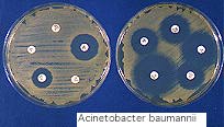
The most novel of these tests were the blood and sputum cultures. Building on the germ theory of disease, the physicians and their associated research scientists attempted to find the culprit for this deadly infection. Physicians would commonly order both blood and sputum cultures of their influenza and pneumonia patients mostly for research and investigative purposes. At the military training camp Camp Lewis during a influenza epidemic, "in all cases of pneumonia...a sputum study, white blood and differential count, blood culture and urine examinations were made as routine," (JAMA, 1/25/1919). The bacterial flora of the nasopharynx of some patients was also cultured since droplet infection was where the disease disseminated. The collected swabs and specimens were inoculated onto blood agar of petri dishes. The grown up bacterial colonies were closely studied to find the causal organism. Commonly found were pneumococcus, streptococcus, staphylococcus and Bacillus influenzae (JAMA, 4/12/1919). These new laboratory tests used in the clinical setting brought in a solid scientific, biological link to the practice of medicine. Medicine had become fully scientific and technologic in its understanding and characterization of the influenza epidemic.
Treatment and Therapy
The therapeutic remedies for influenza patients varied from the newly developed drugs to oils and herbs. The therapy was much less scientific than the diagnostics , as the drugs had no clear explanatory theory of action. The treatment was largely symptomatic, aiming to reduce fever or pain. Aspirin, or acetylsalicylic acid was a common remedy. For secondary pneumonia doses of epinephrin were given. To combat the cyanosis physicians gave oxygen by mask or some injected it under the skin (JAMA, 10/3/1918). Others used salicin which reduced pain, discomfort and fever and claimed to reduce the infectivity of the patient. Another popular remedy was cinnamon in powder or oil form with milk to reduce temperature (BMJ, 10/19/1918). Finally, salt of quinine was suggested as a treatment. Most physicians agreed that the patient should be kept in bed (BMJ, 7/13/1918). With that was the advice of plenty of fluids and nourishment. The application of cold to the head, with warm packs or warm drinks was also advised. Warm baths were used as a hydrotherapeutic method in hospitals but were discarded for lack of success (JAMA, 10/3/1918). These treatments, like the suggested prophylactic measures of the public health officials, seemed to originate in the common social practices and not in the growing field of scientific medicine. It seems that as science was entering the medical field, it served only for explanatory, diagnostic and preventative measures such as vaccines and technical tests. This science had little use once a person was ill.
However, a few proposed treatment did incorporate scientific ideas of germ theory and the immune system. O'Malley and Hartman suggested to treat influenza patients with the serum of convalescent patients. They utilize the theorized antibodies to boost the immune system of sick patients. Other treatments were "digitalis," the administration of isotonic glucose and sodium bicarbonate intravenously which was done in military camps (JAMA, 1/4/1919). Ross and Hund too utilized ideas about the immune system and properties of the blood to neutralize toxins and circulate white blood cells. They believed that the best treatment for influenza should aim to: "...neutralize or render the intoxicant inert...and prevent the blood destruction wit its destructive leukopenia and lessened coagulability," (JAMA, 3/1/1919). They tried to create a therapeutic immune serum to fight infection. These therapies built on current scientific ideas and represented the highest biomedical, technological treatment like the antitoxin to diphtheria.
The Etiology of Influenza in 1918
During the 1890 influenza epidemic, Pfeiffer found what he determined to be the microbial agent to cause influenza. In the sputum and respiratory tract of influenza patients in 1892, he isolated the bacteria Bacillus influenzae , which was accepted as the true "virus" though it was not found in localized outbreaks (BMJ, 11/2/1918). However, in studies of the 1907-8 epidemic in the US, Lord had found the bacillus in only 3 of 20 cases. He also found the bacillus in 30% of cultures of sputum from TB patients. Rosenthal further refuted the finding when he found the bacillus in 1 of 6 healthy people in 1900 (JAMA, 1/18/1919). The bacillus was also found to be present in all cases of whooping cough and many cases of measles, chronic bronchitis and scarlet fever (JAMA, 10/5/1918). The influenza pandemic provided scientists the opportunity to confirm or refute this contested microbe as the cause of influenza. The sputum studies from the Camp Lewis epidemic found only a few influenza cases harvesting the influenza bacilli and mostly type IV pneumococcus . They concluded that "the recent epidemic at Camp Lewis was an acute respiratory infection and not an epidemic due to Bacillus influenzae ," (JAMA, 1/25/1919). This finding along with others suggested to most scientists that the Pfeiffer's Bacillus was not the cause of influenza.
In the 1918-19 influenza pandemic, there was a great drive to find the etiological agent responsible for the deadly scourge. Scientists in their labs were working hard, using the cultures obtained from physician clinics, to isolate the etiological agent for influenza. As a report early in the epidemic said, "the 'influence' of influenza is still veiled in mystery, " (JAMA, 10/5/1918). The nominated bacillus influenzae bacteria seemed to be incorrect and scientists scrambled to isolate the true cause. In the journals, many authors speculated on the type of agent-- was it a new microbe, was it a bacteria, was it a virus? One journal offered that "the severity of the present pandemic, the suddenness of onset...led to the suggestion that the disease cannot be influenza but some other and more lethal infection," (BMJ, 11/2/1918). However, most accepted that the epidemic disease was influenza based on the familiar symptoms and known pattern of disease. The respiratory disease of influenza was understood to give warning in the late spring of its potential effects upon its recrudescence once the weather turned cold in the winter (BMJ, 10/19/1918). One article with foresight stated that "there can be no question that the virus of influenza is a living organism...it is possibly beyond the range of microscopic vision," (BMJ, 11/16/1918). Another article confirmed the idea of an "undiscovered virus" and noted that pneumococccci and streptococci were responsible for "the gravity of the secondary pulmonary complications," (BMJ, 11/2/1918). The article went on to offer the idea of a symbiosis of virus and secondary bacterial infection combining to make it such a severe disease.
The investigators as they attempted to find the responsible agent for the influenza pandemic were developing ideas of infectious microbes and the concept of the virus. The idea of the virus as an infectious agent had been around for years. The articles of the period refer to the "virus" in their discussion but do not consistently use it to be an infectious microbe, distinctive from bacteria. The term virus has the same usage and application as bacillus. In 1918, a virus was defined scientifically to be a submicroscopic infectious entity which could be filtered but not grown in vitro . In the 1880s Pasteur developed an attenuated vaccine for the rabies virus by serial passage way ahead of his time. Ivanoski's work on the tobacco mosaic virus in 1890s lead to the discovery of the virus. He found an infectious agent that acted as a micro organism as it multiplied yet which passed through the sterilizing filter as a nonmicrobe. By the 1910s several viruses, defined as filterable infectious microbes, had been identified as causing infectious disease (Hughes). However, the scientists were still conceptually behind in defining a virus; they distinguished it only by size from a bacteria and not as an obligate parasite with a distinct life cycle dependent on infecting a host cell.

The influenza epidemic afforded the opportunity to research the etiological agent and develop the idea of the virus. Experiments by Nicolle and Le Bailly in Paris were the earliest suggestions that influenza was caused by a "filter-passing virus," (BMJ, 11/2/1918). They filtered out the bacteria from bronchial expectoration of an influenza patient and injected the filtrate into the eyes and nose of two monkeys. The monkeys developed a fever and a marked depression. The filtration was later administered to a volunteer subcutaneously who developed typical signs of influenza. They reasoned that the inoculated person developed influenza from the filtrate since no one else in their quarters developed influenza (JAMA, 12/28/1918). These scientists followed Koch's postulates as they isolated the causal agent from patients with the illness and used it to reproduce the same illness in animals. Through these studies, the scientists proved that influenza was due to a submicroscopic infectious agent and not a bacteria, refuting the claims of Pfeiffer and advancing virology. They were on their way to discerning the virus and characterizing the orthomyxo viruses that lead to the disease of influenza.

These scientific experiments which unravel the cause of influenza, had immediate preventative applications. They would assist in the effort to create a effective vaccine to prevent influenza. This was the ultimate goal of most studies, since vaccines were thought to be the best preventative solution in the early 20th century. Several experiments attempted to produce vaccines, each with a different understanding of the etiology of fatal influenza infection. A Dr. Rosenow invented a vaccine to target the multiple bacterial agents involved from the serum of patients. He aimed to raise the immunity to against the bacteria, the "common causes of death," and not the cause of the initial symptoms by inoculating with the proportions found in the lungs and sputum (JAMA, 1/4/1919). The vaccines made for the British forces took a similar approach and were "mixed vaccines" of pneumococcus and lethal streptococcus. The vaccine development therefore focused on the culture results of what could be isolated from the sickest patients and lagged behind the scientific progress.


